


Syringoma removal in Suwon: how is it different from milia under the eyes, and why is it treated in stages?
If small grain-like bumps rise under your eyes and cannot be covered even with makeup, it is worth first checking whether they are syringoma or milia. Syringoma is a benign bump formed when the fine ducts through which sweat exits enlarge in the dermis, and milia are white granules of dead skin cells that could not shed and became trapped in a shallow part of the skin.
Even though the two lesions look similar, they sit at different depths, so the treatment differs. Milia are usually resolved in one go by making a small opening and removing the contents, while syringoma has roots extending into the dermis, so it is treated over several sessions with a CO2 laser, dividing the depth. At our Suwon clinic, during consultation we first look at the type and number of bumps, and if the two lesions are mixed, we separate which will be handled and how before deciding on the treatment range.
In this article, we outline in order why syringoma and milia form, how to tell under-eye bumps apart, why laser treatment is divided into stages and the process, recovery and recurrence management, and side effects.
Syringoma: key points
- Syringoma is an enlargement of sweat duct tissue in the dermis, and milia are granules of dead skin cells trapped in a shallow layer.
- Syringoma appears as flat, skin-colored bumps clustered symmetrically under both eyes, while milia are one or two white granules scattered apart.
- Because under-eye skin is thin, syringoma treatment continues at 1- to 3-month intervals with a CO2 laser, dividing the depth.
- Red marks after treatment are part of the healing process and fade over 1–2 months.
Why do syringoma and milia form?
Syringoma forms as the tissue around the ducts that run from the sweat glands to the skin surface enlarges. It tends to appear under and around the eyes in women after puberty, and if a family member has similar bumps, they may appear at a similar age. The exact cause has not yet been identified, and the number tends to increase little by little with age.
Milia are a state in which the skin's process of shedding dead cells is blocked and clumps of keratin become trapped in small pockets. They can form without any particular trigger, or during the healing of burns, laser treatments, or scratches. They tend to appear where the skin is thin, such as around the eyes, and some disappear on their own over time.
Syringoma rarely disappears on its own. Since there is almost no pain or itching, it is easy to leave alone, but as the number increases, the range to treat at once widens and the course lengthens.
How do you tell syringoma from milia under the eyes?
They differ in the visible color and shape, how they cluster, and how they feel to the touch. The table below shows the criteria we use to distinguish the two lesions during consultation.
| Category | Syringoma | Milia |
|---|---|---|
| Cause | Enlargement of sweat duct tissue in the dermis | Keratin trapped in a shallow layer |
| Color | Skin-colored or pale yellowish | Appears white beneath the skin |
| Size and shape | Flat, firm bumps of 2 to 3 mm | Round granules of 1 to 2 mm |
| Distribution | Several clustered symmetrically under both eyes | One or two scattered apart |
| Depth | Extends into the dermis | Shallow layer of the skin |
If you squeeze by hand, the firm granule of milia does not come out easily, and syringoma has no contents to come out. Squeezing can cause a wound that leaves pigmentation, so it is best not to touch them at home. Lesions that look similar, such as flat warts or xanthomas, also occur under the eyes, so if the appearance is unclear, the medical team examines them under magnification to tell them apart.
Why is syringoma removal done over several sessions?
How CO2 laser removal works
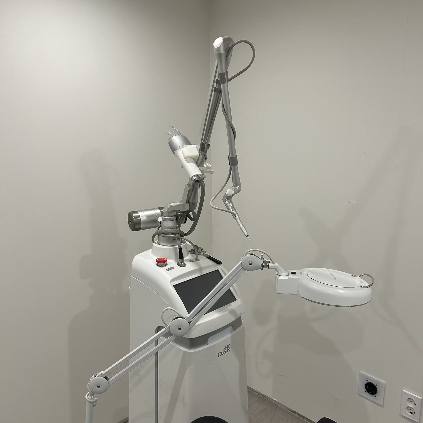
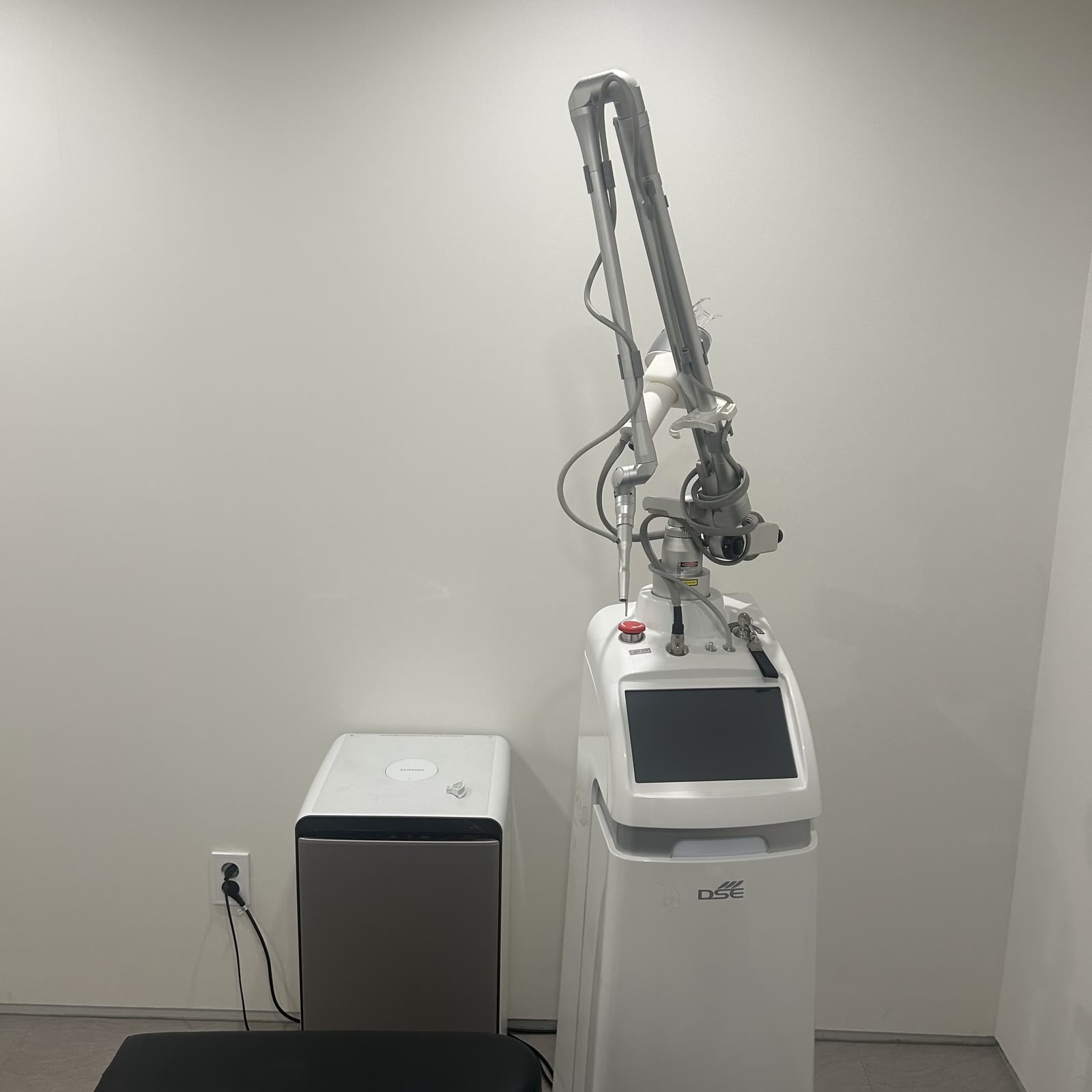
A CO2 laser vaporizes lesion tissue using heat generated as light at a wavelength of 10,600nm is absorbed by water in the skin. Shallow lesions such as milia can be emptied just by opening the entrance, but with syringoma, the enlarged duct tissue in the dermis must also be addressed to reduce the chance of it coming back.
Under-eye skin is among the thinnest on the face, so shaving down to the depth of the roots at once increases the risk of pitted scars or pigmentation. That is why at each session we remove only to the depth the skin can tolerate, and after it heals, we address the remaining tissue again. The number of sessions is decided based on the number and depth of the bumps, how widely they have spread, and previous treatment history.
Treatment steps at our Suwon clinic
- Personalized Consultation
- Photos (Before Treatment)
- Numbing Cream
- Treatment
- Photos (After Treatment)
- Finishing Care
Anesthesia is done with numbing cream, and the treatment takes about 1 hour or less, though it varies with the number and range of bumps. The treatment range is set by dividing the whole face and the whole neck each into 50 or fewer, 100 or fewer, and more than 100. The next session is scheduled at 1- to 3-month intervals after the treated area has fully healed, and we compare remaining bumps using photos taken before and after the treatment.
View CO2 laser treatment information for moles, milia, and syringoma removal
Recovery and recurrence management after syringoma removal
- Don't pick at the scab by hand — let it fall off on its own.
- Red marks where the scabs have fallen off are a reaction that occurs during healing and fade over 1–2 months.
- For 3–7 days after treatment, avoid heavy drinking, smoking, saunas and jjimjilbang, hot baths, and strenuous exercise to prevent inflammation.
- Depending on the treated area, medication is prescribed to prevent inflammation and relieve pain; take it for 3 days without missing a dose.
- While red marks remain, apply sunscreen every time you go out to reduce pigmentation.
Tissue remaining deep down may rise again, or new bumps may appear nearby. It is hard to expect everything to disappear with a single treatment, so between sessions we check the remaining parts and re-treat only the spots that need it. If you notice any new bumps before your next visit, please let us know.
Side effects and precautions after laser removal
Right after the treatment, stinging, redness, mild swelling, and small scabs appear, and most are reactions that occur during healing. After the scabs fall off, red or light brown marks may remain for a while, and the speed of recovery and the results vary from person to person.
Rarely, long-lasting pigmentation, slightly pitted scars, or hypopigmentation where color turns white may occur. These risks increase if you forcibly pick off scabs or get a lot of sun exposure during recovery, and those prone to keloids, where wounds heal thickly, are also at greater risk. If you have a medical history such as an antibiotic allergy, are pregnant or breastfeeding, or are taking medication that makes you sensitive to light, please be sure to tell us before the treatment.
If oozing continues, swelling and heat do not decrease but worsen after several days, or you see yellow pus, these may be signs of inflammation, so please contact the clinic right away.
What should I check before syringoma removal?
If any of these apply, get a diagnosis first
- Flat, skin-colored bumps are clustered symmetrically under both eyes.
- Nothing comes out when I squeeze them, and they feel firm to the touch.
- The number of bumps has gradually increased over the past few years.
- A family member has similar bumps under their eyes.
If two or more apply, it is best to first check what kind of bumps they are before laser treatment. Whether to proceed, the method, and the scope are decided together in consultation after the medical team examines you.
In short, the key with syringoma is to first distinguish it from milia, since they lie at different depths, and to treat it in stages matched to the pace at which the skin heals.
FAQ
Q. Can syringoma be removed with ointment or medication?
A. Because it is a lesion in which duct tissue in the dermis has enlarged, topical medication can hardly reach that depth. That is why we use a method that removes the tissue directly, such as a CO2 laser, and proceed over several sessions matched to the depth of the bumps.
Q. Can I squeeze milia at home?
A. Because the surface of milia is closed, they do not come out easily by hand, and forcing them can leave wounds and pigmentation. Making a small opening with a sterilized instrument and then removing the contents is easier on the skin, and if they are mixed with syringoma, we first separate them through an examination.
Q. About how many sessions does under-eye syringoma take?
A. It depends on the number and depth of the bumps and how widely they have spread, so the number of sessions is decided in consultation. After confirming that the treated area has fully healed, we continue at 1- to 3-month intervals and compare remaining bumps with photos at each session.
Q. Can treated syringoma come back?
A. Tissue remaining deep down may rise again, or new bumps may appear nearby. That is why between sessions we check the remaining parts and re-treat only the spots that need it, and if you notice any new bumps, just let us know at your next visit.
Q. When can I wear makeup after laser removal?
A. Until the scabs fall off, it is best to keep cosmetics off the treated area and not rub it. When you can start wearing makeup again depends on the treatment range and how quickly the scabs fall off, so we give guidance for each area after the treatment.
Q. How can I find out the cost of syringoma removal?
A. The cost depends on the number of bumps to be removed, the area, and the number of sessions. We guide you after confirming in consultation how many bumps on the face or neck will be treated, and you can also check the items by treatment area on the treatment information page.
This content is provided as general treatment information and is not a substitute for diagnosis or treatment. Always consult a medical professional to decide whether and how to proceed with a treatment.