


Milia removal in Suwon: why you shouldn't squeeze white bumps around the eyes
If white, grain-like bumps have appeared around your eyes and bother you because they won't come out when pressed, it is worth checking for milia before assuming acne. Milia are white bumps 1–2 mm in size formed when dead skin cells that should shed off the skin become trapped and hardened under thin skin, with a thin layer of cells wrapping the keratin clump like a pouch.
Milia are not structured to come out when squeezed, so they are removed by making a very small opening on the surface and extracting the bump along with its pouch. At our Suwon clinic, we first check the location and number of bumps during the consultation, and whether they are mixed with syringomas or skin tags, before deciding the area to treat with the CO2 laser.
In this article, we go over why milia form and why they won't come out when squeezed, how to tell them apart from closed comedones and syringomas, the milia removal process, recovery care, and side effects, in that order.
Milia: key points
- Milia are keratin trapped and hardened in a pouch under the skin, and since there is no passage for it to exit, they won't come out when pressed.
- Closed comedones release their contents when squeezed, and syringomas differ from milia in that several skin-colored bumps are spread under the eyes.
- Milia are removed either by making a small opening on the surface to extract the bump, or by vaporizing it along with the surrounding layer using a CO2 laser.
- Red marks after treatment are part of the healing process and fade over 1–2 months.
Why do milia form, and why won't they come out when squeezed?
Milia are divided into two types depending on how they form. Primary milia, which form without any particular trigger, can appear at any age from infants to adults and are commonly seen on the eyelids, under the eyes, and on the cheeks. Secondary milia form where the skin was once injured and then covered over again, such as healed burns or blisters, or areas that have recovered after peels or laser treatments, because keratin cannot escape.
They form especially often around the eyes because the skin there is thin and dead skin cells tend to shed unevenly. Repeated habits such as applying heavy eye cream thickly or going to sleep without fully removing eye makeup make keratin more likely to build up. Since they are not caused by bacteria or viruses, they do not spread to other people.
Milia have no passage for sebum to exit, so pressing them by hand leaves the bump in place and only crushes the surrounding skin. Because capillaries are close to the surface around the eyes, pressing just a few times can cause bruising, and brown pigmentation may remain there for a long time. Picking them with a needle at home can easily let bacteria in and cause inflammation, and if only part of the contents comes out and the pouch remains, it fills up again in the same spot.
How do you tell milia apart from closed comedones and syringomas?
Small bumps around the eyes look similar and are easy to confuse. Looking at the cause, shape, where they tend to appear, and how they react when pressed can help tell them apart.
| Category | Milia | closed comedones | Syringoma |
|---|---|---|---|
| Cause | Keratin trapped inside a pouch | Sebum and dead skin cells clogging a pore | Enlarged sweat ducts |
| Shape and color | Firm white bump, 1–2 mm | Soft white or skin-colored bump | Flat skin-colored or yellowish bump, 2–3 mm |
| Common locations | Eyelids, under the eyes, cheeks | Forehead, cheeks, chin | Several spread out under the eyes |
| When pressed | Nothing comes out | Sebum plugs may come out | Nothing comes out |
Syringomas are rooted in the deep layers of the skin, so they cannot be cleared by extraction like milia, and the depth treated with the laser is different. Soft, skin-colored bumps that dangle on the eyelids or neck may be skin tags. If there are several similar bumps under the eyes, you need to first determine what kind of lesion they are before a removal method can be chosen.
How is milia removal performed?
How milia removal works
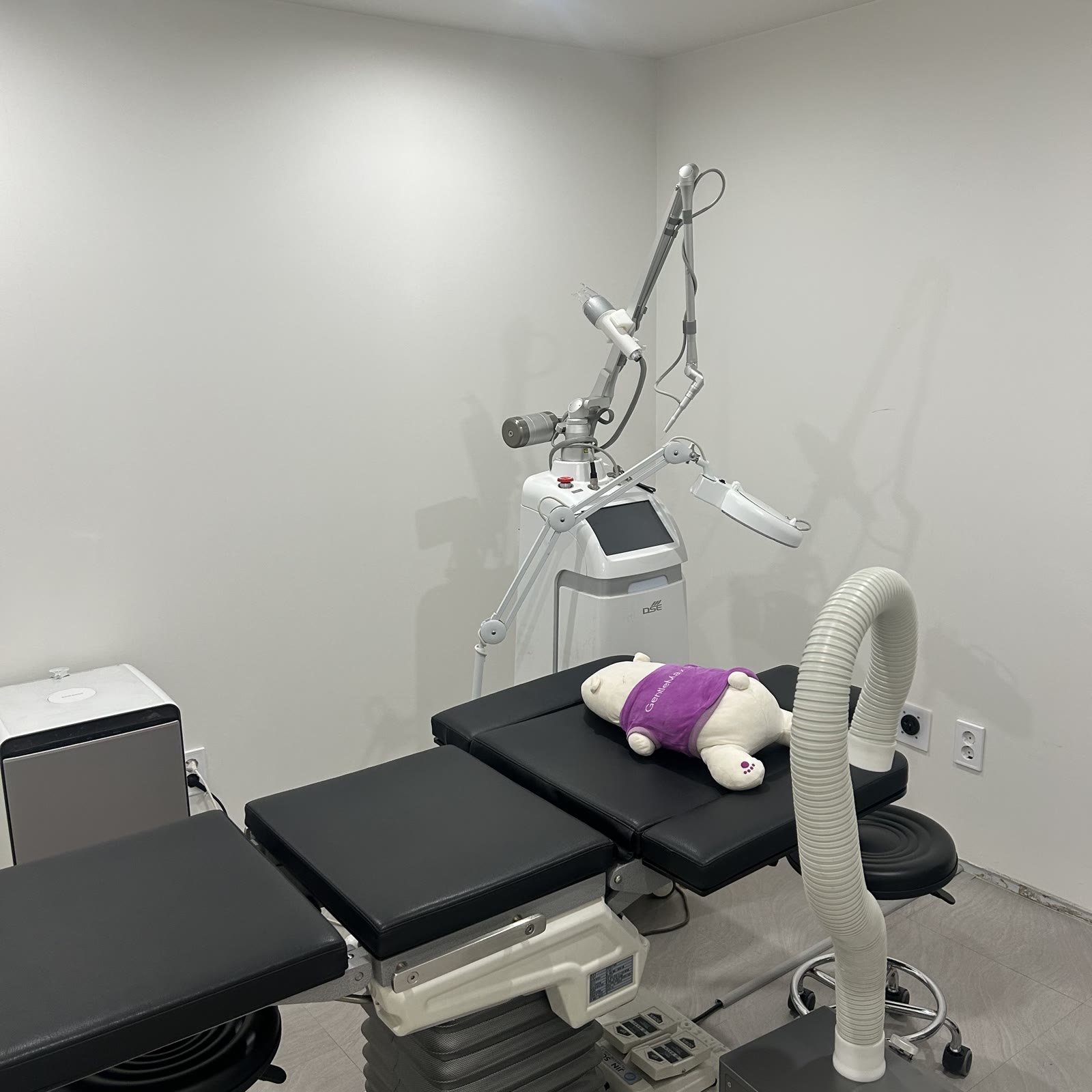
Milia close to the surface can also be removed by making a tiny hole on the surface with a sterilized needle or fine blade tip and pushing out the keratin clump with an extractor. Milia that are clustered together or have reappeared in the same spot are treated with a CO2 laser, which vaporizes the surface and the layer of cells wrapping the bump together.
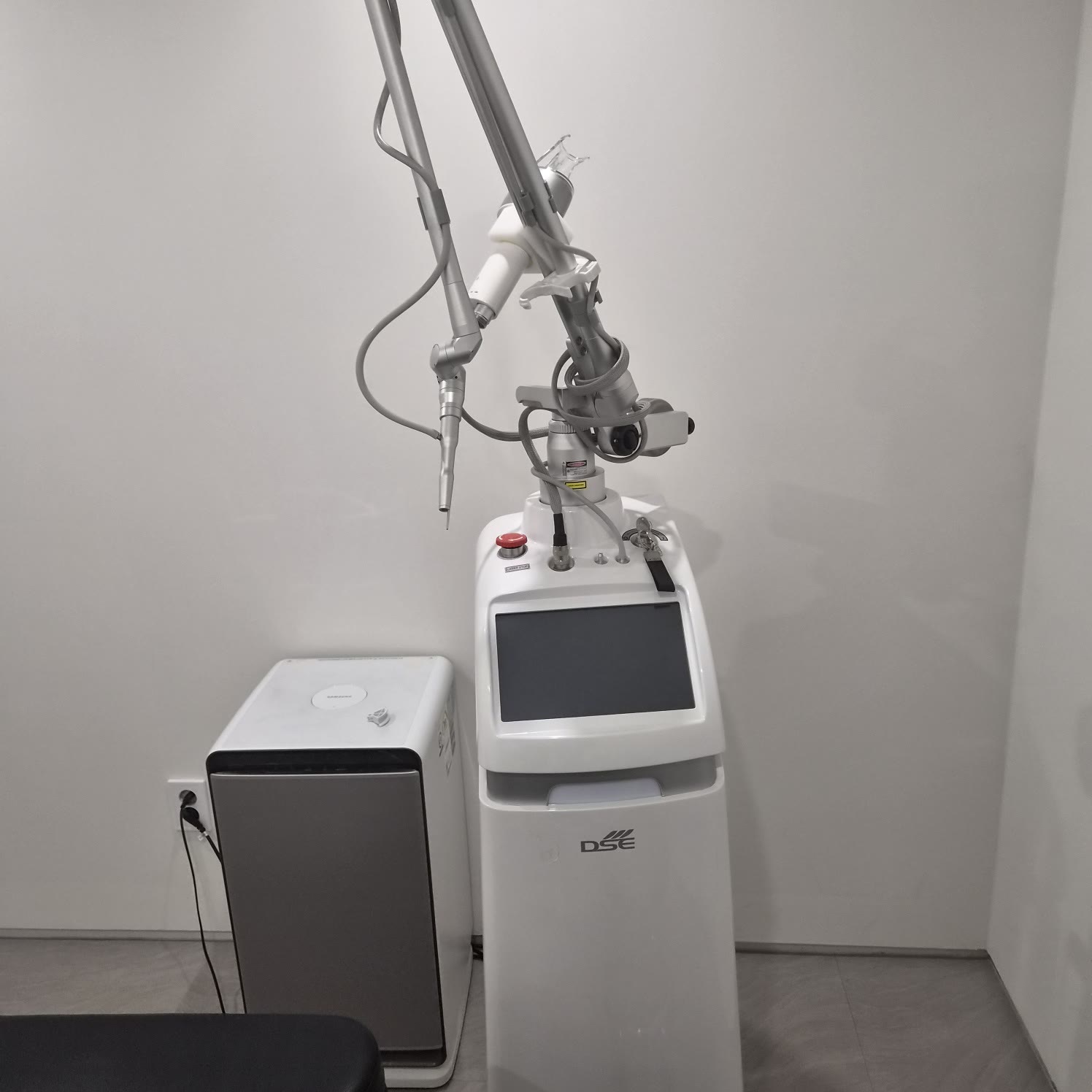
A CO2 laser uses the property of 10,600 nm light being absorbed by water in the skin. Because it can be applied as narrowly as the size of the bump, the area can be kept small even on thin skin like the eyelids, and removing the pouch as well reduces the chance of it filling up again in the same spot.
Treatment steps at our Suwon clinic
- Personalized Consultation
- Photos (Before Treatment)
- Numbing Cream
- Treatment
- Photos (After Treatment)
- Finishing Care
Anesthesia is done with a numbing cream, and the procedure takes about an hour or less, depending on the number of bumps and the extent. The treatment area is set by dividing the whole face and the whole neck, each into tiers of up to 50, up to 100, and over 100. When treating newly formed milia again, we wait 1–3 months until the previously treated areas have fully healed.
View CO2 laser milia removal details
Recovery and recurrence care after milia removal
- Do not pick off or rub the scabs; let them fall off on their own.
- Red marks after treatment are not a side effect but a reaction that occurs during healing, and they fade over 1–2 months.
- For 3–7 days after treatment, avoid heavy drinking, smoking, saunas and jjimjilbang, hot baths, and strenuous exercise to prevent inflammation.
- Depending on the treated area, medication is prescribed to prevent inflammation and relieve pain; take it for 3 days without missing a dose.
- During recovery, apply sunscreen when going out and also wear a hat or sunglasses.
Spots where the pouch has been removed are less likely to fill up again, but if your skin tends to build up keratin, new milia may form in other spots. Thoroughly removing eye makeup and applying eye cream thinly may help reduce new bumps. Avoid frequently using harsh exfoliants or scrubs around the eyes, as they can make the skin more sensitive instead.
Side effects and precautions for milia removal
Right after treatment, small red dots and scabs appear, and most of these are reactions that occur during healing. Because the skin around the eyes is thin, swelling may remain for a day or two, and recovery speed and results vary from person to person.
In rare cases, pigmentation, small depressed marks, or inflammation can occur. Picking off scabs early or getting a lot of UV exposure during recovery increases the risk of pigmentation, and people prone to keloids, where wounds heal thick, are also at higher risk. If you have a medical history such as an antibiotic allergy or any special conditions, be sure to tell us before treatment.
If the treated area becomes increasingly swollen and painful, you see pus, or your eyes become red and your vision feels uncomfortable, please contact the clinic right away.
What should be checked before milia removal?
If any of these apply, get a diagnosis first
- There are white bumps around or under my eyes that won't come out when pressed.
- The bumps have stayed the same for months or have increased in number.
- Bumps have reappeared where I squeezed or picked them with a needle at home.
- Several skin-colored or yellowish bumps are spread out together under my eyes.
If two or more apply, it is a good idea to confirm what kind of lesion it is before squeezing the bumps yourself. Whether to treat, which method to use, and the treatment area are decided together during the consultation after the doctor's examination.
In short, the key with milia is not to squeeze them by hand, to tell them apart from syringomas, to remove them along with the pouch, and to manage the keratin around the eyes so new bumps don't form.
FAQ
Q. Do milia under the eyes go away on their own?
A. Milia in babies sometimes disappear within a few weeks, but milia around the eyes in adults tend to remain for months or longer. If they are getting bigger or increasing in number, we recommend first checking their location and type during a consultation.
Q. Can milia on the eyelids also be removed?
A. Milia on the eyelids can also be removed, but because the skin is thin, the area must be kept small. We check the location and depth of the bumps and their distance from the eye during the examination before deciding how to remove them, and if there are many, treatment may be divided into sessions.
Q. Do milia come back after they are removed?
A. Spots where the pouch has been removed are less likely to fill up again, but if your skin tends to build up keratin, new milia may form in other spots. Thoroughly removing eye makeup and applying eye cream thinly may help.
Q. Does milia removal hurt?
A. A numbing cream is applied to the treatment area and given enough time to work before the procedure, which reduces the stinging sensation. How much you feel depends on the area, the number of bumps, and the individual, and thin-skinned areas such as the eyelids may feel a little more sensitive.
Q. When can I start washing my face and wearing makeup after milia removal?
A. Until the scabs fall off, do not rub the treated area; wash gently with lukewarm water and pat dry. When you can resume makeup depends on the treatment area and how quickly the scabs come off, so we will let you know after the procedure.
Q. How can I find out the cost of milia removal?
A. The cost depends on the number of bumps and the treatment area. We confirm during the consultation how many milia on the face or neck will be treated before giving you the details, and you can also check the items for each treatment area on our treatment information page.
This content is provided as general treatment information and is not a substitute for diagnosis or treatment. Always consult a medical professional to decide whether and how to proceed with a treatment.